
In this storyOpen index
Rosalind Franklin: The Scientist Who Photographed DNA and Died One Day Before Seeing Her Final Discovery
Photograph 51 revealed the hidden architecture of life, but Franklin’s scientific legacy extended far beyond DNA.
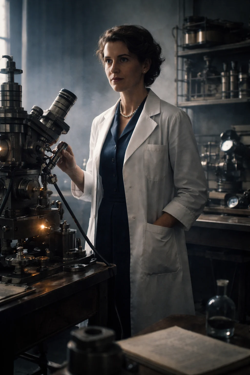
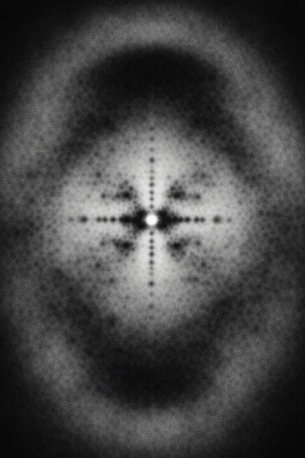
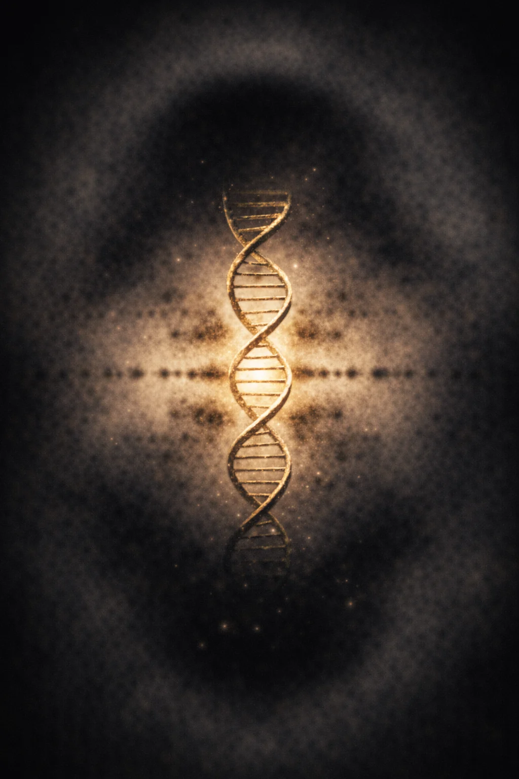
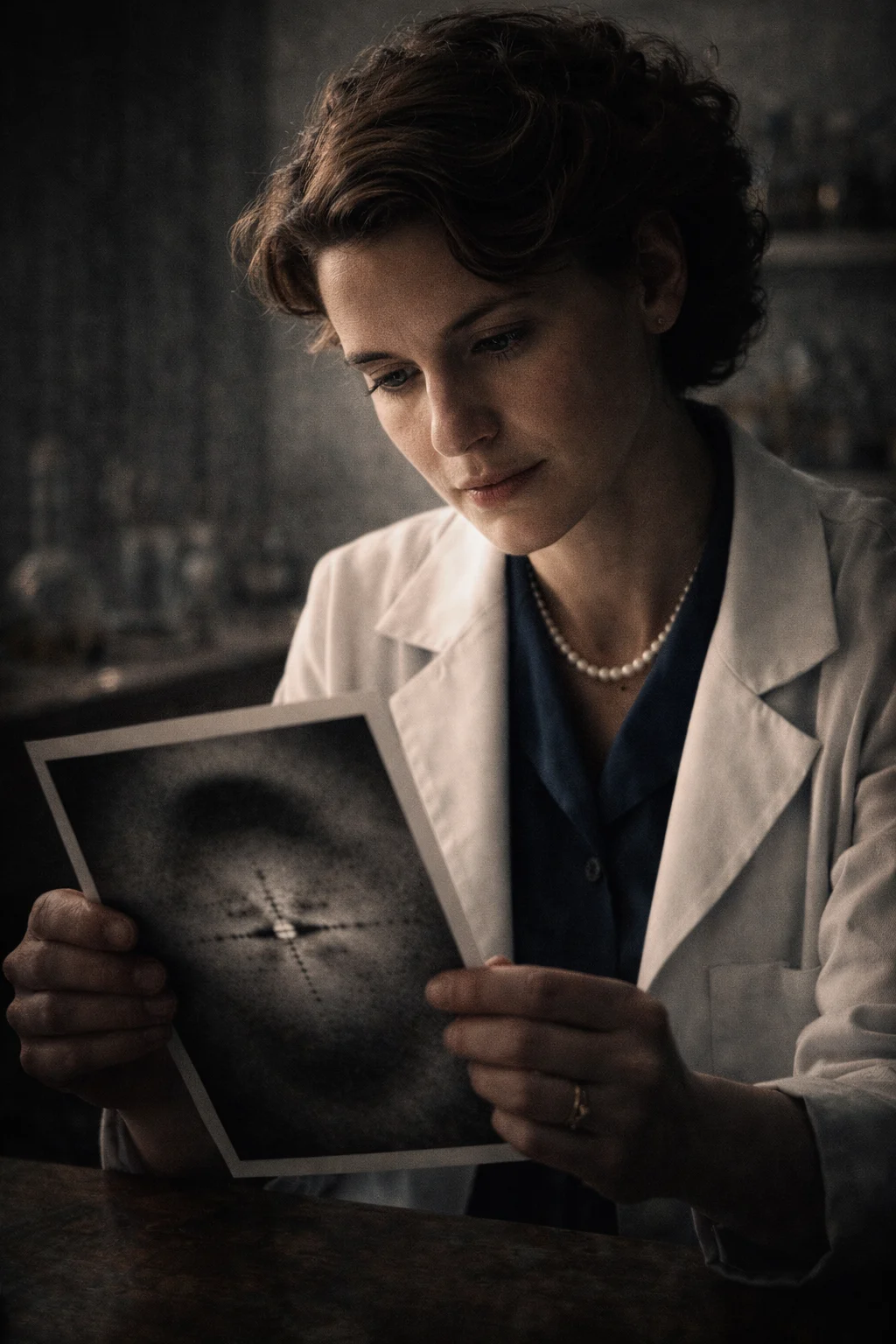
The photograph that transformed biology showed neither a face, a landscape, nor a historic event. It was a dark figure crossed by a pattern of black spots. To almost anyone else, it might have appeared to be a failed image. To Rosalind Franklin, it revealed the hidden architecture of life.
The celebrated Photograph 51 helped demonstrate that DNA possessed a helical structure. Reducing Franklin’s career to that image, however—or presenting her only as a scientist deprived of a prize—conceals much of her true legacy.
Before studying DNA, she investigated the structure of coal. Afterwards, she became an authority on viruses, directed her own research team, and secured significant international funding. Her final project was displayed at the 1958 Brussels World’s Fair. Franklin never saw it: she died the day before the exhibition opened.
Rosalind Franklin on video
Discover this story on our YouTube channel.
A life told through places and turning points

Enter the archive
An authentic object preserved by a cultural institution brings us closer to the world behind this life.
- Collection
- Wikimedia Commons
- Rights
- CC BY-SA 4.0
From the personal collection of Jenifer Glynn.
A girl who wanted demonstrable answers
Rosalind Elsie Franklin was born in London on July 25, 1920, into an affluent Jewish family that valued education and public service. From childhood, she showed an aptitude for mathematics and a strong preference for rational explanations.
At fifteen, she decided to become a scientist. Her father, Ellis Franklin, initially expressed doubts about a career then considered highly unusual for a woman, but he ultimately supported her education.
In 1938, she entered Newnham College, one of the women’s colleges associated with the University of Cambridge. She studied natural sciences and specialized in physical chemistry. Women could study and complete university examinations, but Cambridge still did not award them degrees on equal terms with men. That institutional discrimination did not end until 1948.
Franklin completed her studies during the Second World War. While London endured air raids, she served as an air-raid warden and began research that seemed far removed from genetics: examining the pores inside coal.
The invisible holes in coal
In 1942, Franklin joined the British Coal Utilisation Research Association. Britain depended on coal for its industries, homes, and war effort. Understanding how different types of coal absorbed and released gases had practical applications in fuel production and filtering materials.
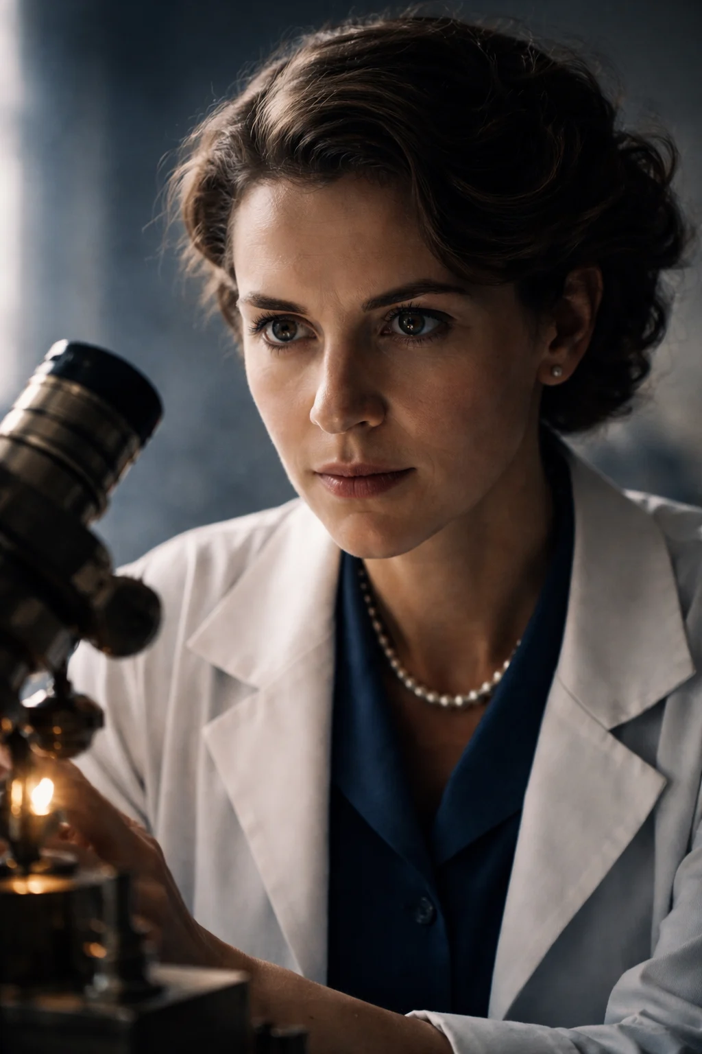
Franklin investigated coal’s density, porosity, and microscopic organization. She demonstrated that its pores formed a complex molecular network and that different materials could be classified according to their behavior when heated. Her research helped predict which coals could be transformed into graphite and which were suitable for particular industrial purposes (National Library of Medicine, n.d.).
This work formed the basis of her doctoral thesis, approved by Cambridge in 1945. It was not an insignificant prelude to her later achievements: her papers on coal and carbon continued to be cited for decades.
In 1947, she moved to Paris to work at the Laboratoire Central des Services Chimiques de l’État. Under Jacques Mering, she mastered X-ray diffraction, a technique used to examine structures too small to be observed through a conventional microscope.
X-rays were directed at a sample and scattered into a pattern. Interpreting the resulting spots demanded experimental precision, mathematical knowledge, and patience. Franklin excelled at all three.
Conflict at King’s College
In 1951, Franklin returned to London to join the biophysics unit at King’s College. Its director, John Randall, assigned her to investigate DNA fibers and placed doctoral student Raymond Gosling under her supervision.
The working arrangement was poorly defined from the beginning. Maurice Wilkins, who had already conducted research on DNA, initially believed that Franklin would be his assistant. She had actually been recruited as an independent researcher. This misunderstanding, combined with their contrasting personalities and the predominantly male culture of King’s, damaged their professional relationship.
Franklin and Gosling discovered that DNA could exist in two forms depending on humidity. The drier A form produced a complicated pattern; the more hydrated B form yielded a much clearer one.
On May 2, 1952, they began an X-ray exposure lasting approximately 62 hours. The result was Photograph 51. Its distinctive dark cross provided strong evidence of a helical structure (King’s College London, 2023).
The photograph alone, however, was not a complete answer. Franklin was attempting to determine the molecule’s dimensions, symmetry, and distribution of components through rigorous calculations. She correctly established that the phosphate groups had to be located on the outside—an essential condition for any valid DNA model.
What really happened to Photograph 51
The story is often condensed into the claim that James Watson and Francis Crick “stole” a photograph and discovered the double helix. The reality was more complicated, though not entirely fair.
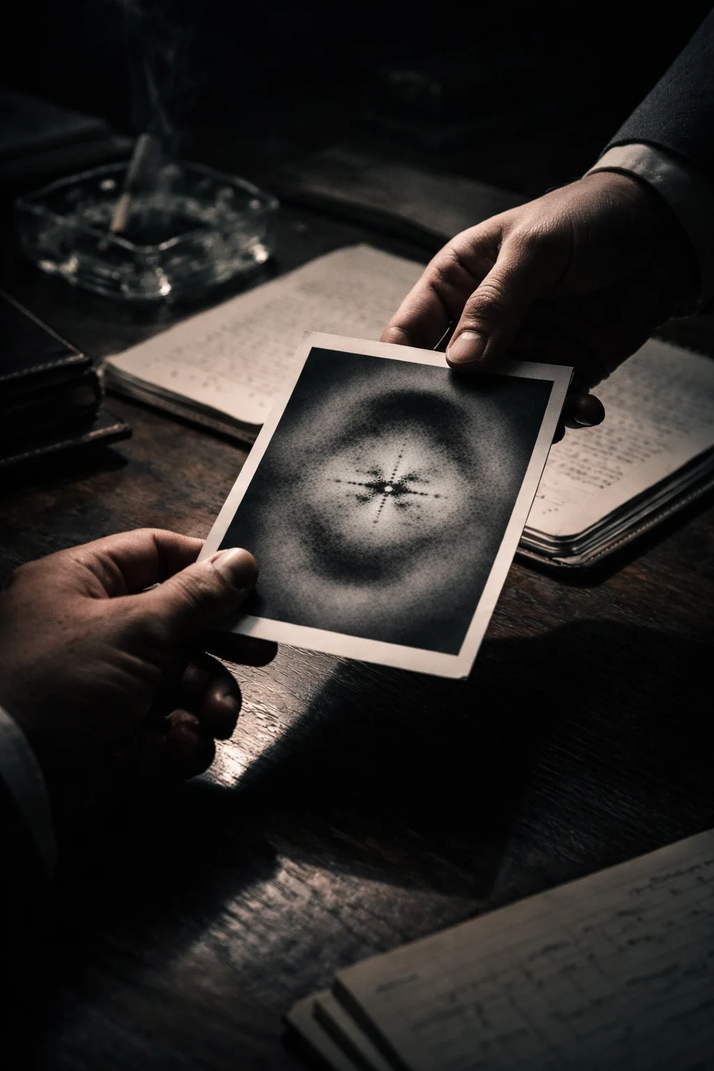
As Franklin prepared to leave King’s, Gosling returned to Wilkins’s supervision. Wilkins showed Photograph 51 to Watson without consulting Franklin. Watson immediately recognized the pattern of a helix.
Max Perutz also gave Watson and Crick an internal Medical Research Council report containing measurements obtained by Franklin. The document was not formally classified as confidential, but she did not know that her results would be used in this way.
These data allowed Watson and Crick to correct their earlier errors and construct the double-helix model. On April 25, 1953, Nature published three consecutive papers: Watson and Crick’s model; the findings of Wilkins, Alexander Stokes, and Herbert Wilson; and Franklin and Gosling’s experimental analysis.
Watson and Crick stated only that they had been “stimulated” by the unpublished results of the King’s researchers. They did not explain the degree to which their model depended on those findings.
Recent historical research has also challenged the portrayal of Franklin as a passive victim who failed to understand her own evidence. Documents from 1953 indicate that she was regarded as a central contributor and that her analysis was moving toward a helical interpretation of DNA (Cobb & Comfort, 2023). She was an active scientist within a collective investigation, although her contribution did not receive proportionate recognition at the time.
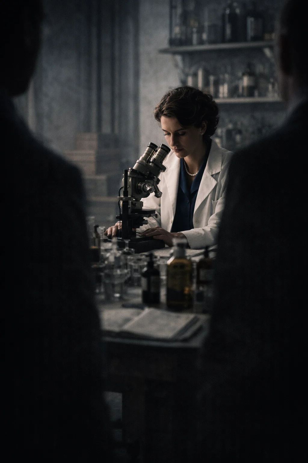
The scientist who continued after DNA
Franklin moved to Birkbeck College in March 1953. Leaving King’s did not end her career. Instead, she began an extraordinary second chapter.
Working under the general direction of John Desmond Bernal, she studied tobacco mosaic virus, which carries RNA rather than DNA. Franklin demonstrated that its particles had a uniform length and helped establish that the RNA was wound along the inside of a helical protein shell.
She was no longer simply a subordinate member of a laboratory. She led a team that included scientists such as Aaron Klug, John Finch, and Kenneth Holmes. Between 1953 and 1958, she published numerous studies on virus structures. Klug continued developing this field and received the 1982 Nobel Prize in Chemistry.
Franklin also secured American funding to expand her research to viruses affecting potatoes, tomatoes, turnips, and peas. Eventually, she began investigating poliovirus, the cause of poliomyelitis.
This period demonstrates that her talent did not depend on a single photograph. After leaving the race to solve DNA, she created her own research program and became a respected figure in structural virology.
The project she never saw
In 1956, just as her team was gaining recognition, Franklin was diagnosed with ovarian cancer. She underwent surgery and treatment but repeatedly returned to her laboratory. During periods of remission, she continued traveling, preparing papers, and seeking funding for her colleagues.
Her group was commissioned to build large virus models for the International Science Pavilion at the 1958 Brussels World’s Fair. Standing approximately five feet tall, the structures allowed members of the public to see how viral particles were organized.
Rosalind Franklin died in London on April 16, 1958. She was thirty-seven. The World’s Fair opened the following day. Visitors were able to examine models created from research she was no longer there to explain (Rosalind Franklin University, n.d.).
Four years later, the Nobel Prize in Physiology or Medicine was awarded to Watson, Crick, and Wilkins for their discoveries concerning the molecular structure of nucleic acids. Franklin could not receive it because Nobel Prizes are not awarded posthumously. Yet saying that the prize was simply “stolen from her” also oversimplifies events. She was never nominated and died before the biological importance of the double helix had been fully confirmed and honored.
More than the woman behind the photograph
For years, Rosalind Franklin was portrayed in two incomplete ways: first as a secondary technician and later as a powerless victim unable to defend her own discovery.
She was much more.
She was a physical chemist with exceptional experimental ability. She produced important research on coal and graphite, obtained fundamental evidence about DNA, and led pioneering investigations into virus structures. She also worked within institutions where hierarchy, misunderstanding, and gender prejudice influenced who received information, authority, and recognition.
Photograph 51 made an invisible structure visible. Franklin’s life reveals something equally difficult to see: scientific discoveries rarely belong to one person, and historical memory does not always distribute credit with the same precision demanded inside a laboratory.
References
- Cobb, M., & Comfort, N. (2023). What Rosalind Franklin truly contributed to the discovery of DNA’s structure. Nature, 616, 657–660.
- Franklin, R. E., & Gosling, R. G. (1953). Molecular configuration in sodium thymonucleate. Nature, 171, 740–741.
- King’s College London. (2023, April 14). The story behind Photograph 51.
- Maddox, B. (2002). Rosalind Franklin: The dark lady of DNA. HarperCollins.
- National Library of Medicine. (n.d.). The Rosalind Franklin papers: Biographical overview.
- National Library of Medicine. (n.d.). The holes in coal: Research at BCURA and in Paris, 1942–1951.
- Nobel Prize Outreach. (n.d.). The Nobel Prize in Physiology or Medicine 1962.
- Rosalind Franklin Institute. (n.d.). Rosalind Franklin’s life.
- Rosalind Franklin University of Medicine and Science. (n.d.). Dr. Rosalind Franklin.
More from this collection
Nicolaus Copernicus: The Man Who Moved the Earth Without a Telescope
Read story →
Isaac Newton: The Man Who Turned the Universe’s Invisible Forces Into Mathematical Laws
Read story →